On this page
I still remember a nine-month-old Bernese Mountain Dog named Otto who came into my treatment room because his owner noticed he’d started “bunny-hopping” up the porch steps instead of taking them one at a time. He weighed close to 70 pounds already, and the front-leg lameness was worse after his morning nap and better once he’d been moving for ten minutes — a pattern that, in my experience, points hard toward the elbow rather than the shoulder. Radiographs confirmed fragmented medial coronoid process on the right side. His owners had assumed a growth spurt was just clumsy. It wasn’t. It was a joint that had grown incongruent with itself.
That’s elbow dysplasia in a sentence: not one disease but a family of developmental problems inside the elbow that keep the three bones of that joint — the humerus, radius, and ulna — from fitting together the way they should as a young dog grows.
What’s Actually Malformed Inside the Joint
The canine elbow is a hinge built from three bones that have to grow at matched rates and align within a narrow tolerance for the joint surfaces to glide smoothly. Elbow dysplasia is the umbrella term veterinarians use for a cluster of developmental lesions that show up when that growth doesn’t stay in sync: fragmented medial coronoid process (a piece of the ulna chips or fails to fuse properly), osteochondritis dissecans (a flap of cartilage separates from the underlying bone), ununited anconeal process (a growth center on the ulna never fuses to the rest of the bone), and elbow incongruity (the joint surfaces are simply mismatched in shape or angle). Any one of these — or more than one at once — creates abnormal, concentrated pressure on cartilage that isn’t built to take it, and that mechanical mismatch is what eventually produces secondary osteoarthritis.
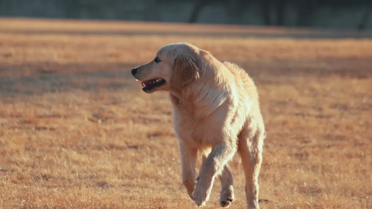
Large and giant breeds are overrepresented because their growth plates are doing more work, faster, over a shorter runway: Labradors, Golden Retrievers, Bernese Mountain Dogs, Rottweilers, and German Shepherds are the breeds I see most often, typically between four and twelve months of age when it first becomes visible.
Reading the Early Signs Correctly
Owners rarely bring a dog in saying “I think it’s the elbow.” They say the dog is stiff after resting, reluctant to go down the stairs, or holding a front leg out slightly when standing — what vets call an abducted or “toed-out” stance that lets the dog unload pressure off the inside of the joint. It’s a subtle limp, not a dramatic one, and that’s exactly why it gets missed for months.
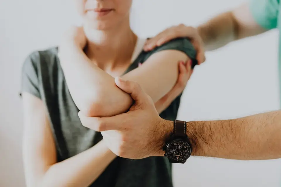
Diagnosis usually starts with a careful orthopedic exam — palpating for pain on elbow flexion and effusion (fluid) in the joint — followed by radiographs, and often CT when the lesion is subtle or surgical planning is on the table, because plain X-rays can miss smaller coronoid fragments.
What Actually Slows the Downstream Damage
Once cartilage has been chipped or overloaded, the joint is on a path toward osteoarthritis, and nothing reverses that structural change. What the evidence supports is slowing the inflammatory and mechanical cascade that follows. Glucosamine and chondroitin remain among the most studied joint-support ingredients for dogs generally, and current research gives each a distinct, modest job rather than a miracle one. A 2026 review in Frontiers in Veterinary Science describes chondroitin as supporting healthy synovial fluid homeostasis — in plain terms, helping maintain the lubricating fluid that lets joint surfaces glide rather than grind. A separate review in the Journal of Veterinary Medical Science found that glucosamine HCl can help prevent experimentally induced cartilage degradation in vitro — a laboratory finding about cartilage cells, worth taking seriously, but not the same thing as a guarantee inside a living, weight-bearing joint. Green-lipped mussel has its own separate literature: a study in Nutrients found that mussel-derived compounds alleviated paw swelling and reduced inflammatory markers including TNF-α and IL-17 in a rat model — again, structure/function-level evidence about inflammation control, not a cure for a malformed joint.
I think of these ingredients the way I think of acupuncture or food therapy in my own practice — each one is a modality that may help support a specific piece of the picture, not a replacement for the piece it can’t touch. Glucosamine can support cartilage matrix maintenance. Chondroitin can support synovial fluid quality. Neither one reshapes a fragmented coronoid process or fuses an ununited anconeal process; only surgery, and sometimes just time and weight management, address the mechanical lesion itself.
Weighing Surgery Against Management — A Real Case
A client of mine had a seven-month-old Golden named Daisy, 58 pounds, whose fragmented medial coronoid process was caught early on radiographs taken because her owner noticed she was slower getting up from her bed in the mornings — not lame exactly, just deliberate about it, and worse on cold days. The surgeon recommended arthroscopic removal of the fragment. The owners weighed it against conservative management: weight control, controlled exercise, and joint-support supplementation, because the cost and recovery time of surgery mattered to their household budget and Daisy’s temperament made a strict rest period genuinely hard. They chose surgery. Eight months later, at her recheck, Daisy was climbing stairs normally again and had lost the toe-out stance — one dog, one outcome, not a promise of what every fragment case will do, but it’s the kind of tradeoff conversation I have weekly.
That case is a reminder that the label “elbow dysplasia” doesn’t tell you which lesion, how severe, or which path fits your dog and your household — only imaging and an orthopedic exam can answer that.
Where Nutrition Realistically Fits
If you’re reading label claims on a joint supplement while your dog is mid-diagnosis, the honest framing is this: these ingredients are structure/function support, not treatment for the malformation itself, and no reputable label should claim otherwise. For a walkthrough of how to actually read a joint-supplement label and check whether the dose on the front of the bag matches anything backed by research, our team’s testing process breaks down what we check before we trust a claim at all.
Quick Recap
Elbow dysplasia isn’t one diagnosis — it’s a name for several possible developmental faults inside a fast-growing joint, most common in large-breed puppies between four and twelve months old. Imaging confirms which lesion is present; surgery, weight management, and controlled exercise address the mechanical problem; joint-support ingredients like glucosamine, chondroitin, and green-lipped mussel have real, specific, modest evidence behind them for supporting cartilage and synovial fluid — not for undoing a malformed joint.
Frequently asked questions
At what age do dogs usually show signs of elbow dysplasia?
Most cases become noticeable between four and twelve months of age, during the period of rapid skeletal growth in large and giant breeds.
Can elbow dysplasia be cured with supplements alone?
No. Supplements like glucosamine, chondroitin, and green-lipped mussel may help support cartilage and joint fluid, but they don't correct the underlying developmental lesion — that requires veterinary diagnosis and, often, surgery.
Is elbow dysplasia the same thing as hip dysplasia?
They're related developmental joint conditions but affect different joints with different anatomy; a dog can have one, both, or neither.
Which breeds are most at risk?
Labrador Retrievers, Golden Retrievers, Bernese Mountain Dogs, Rottweilers, and German Shepherds are among the most commonly affected large breeds.
Sources
- Chondroitin sulfate and synovial fluid homeostasis review — Frontiers in Veterinary Science
- Glucosamine HCl and cartilage degradation review — Journal of Veterinary Medical Science
- Blue mussel oil compounds and inflammatory markers study — Nutrients